Kashif Igbal1,3 ![]() ,
Qaiser Jamal4,
Javeid Igbal1,
Maria Sadaf Afreen5,
Muhammad Zeeshan Ahmed Sandhu2,
Eshwa Dar2,
Umar Farooq2,
Mohammad Fahd Mushtaq2,
Numera Arshad2,
Muhammad Mohsin Iqbal2
,
Qaiser Jamal4,
Javeid Igbal1,
Maria Sadaf Afreen5,
Muhammad Zeeshan Ahmed Sandhu2,
Eshwa Dar2,
Umar Farooq2,
Mohammad Fahd Mushtaq2,
Numera Arshad2,
Muhammad Mohsin Iqbal2
For correspondence:- Kashif Igbal Email: kashifiqbal321@gmail.com Tel:+923356951284
Received: 1 November 2016 Accepted: 16 January 2017 Published: 26 February 2017
Citation: Igbal K, Jamal Q, Igbal J, Afreen MS, Sandhu MZ, Dar E, et al. Luteolin as a potent anti-leishmanial agent against intracellular Leishmania tropica parasite. Trop J Pharm Res 2017; 16(2):337-342 doi: 10.4314/tjpr.v16i2.11
© 2017 The authors.
This is an Open Access article that uses a funding model which does not charge readers or their institutions for access and distributed under the terms of the Creative Commons Attribution License (http://creativecommons.org/licenses/by/4.0) and the Budapest Open Access Initiative (http://www.budapestopenaccessinitiative.org/read), which permit unrestricted use, distribution, and reproduction in any medium, provided the original work is properly credited..
Purpose: To examine the anti-leishmanial and cytotoxic effects of five naturally occurring phenolic compounds: luteolin (1), lalioside (2), luteolin-4’-O-β-D-glucopyranoside (3), apigenin 4-O-β-D-glucopyranoside (4) and apigenin (5) on Leishmania tropica KWH23 amastigotes .
Methods: The compounds were isolated from the leaves of Lawsonia Inermis via hyphenated high performance liquid chromatography-high resolution mass spectrometry coupled with solid phase extraction-tube transfer nuclear magnetic resonance technique. The isolated compounds were given intraperitoneally to L. tropica KWH23 amastigotes-infected albino mice at a dose of ≥ 3 mg/kg for 5 days. Amphotericin-B was used as standard (reference) drug. Lymphocytes were used to analyze their cytotoxicity.
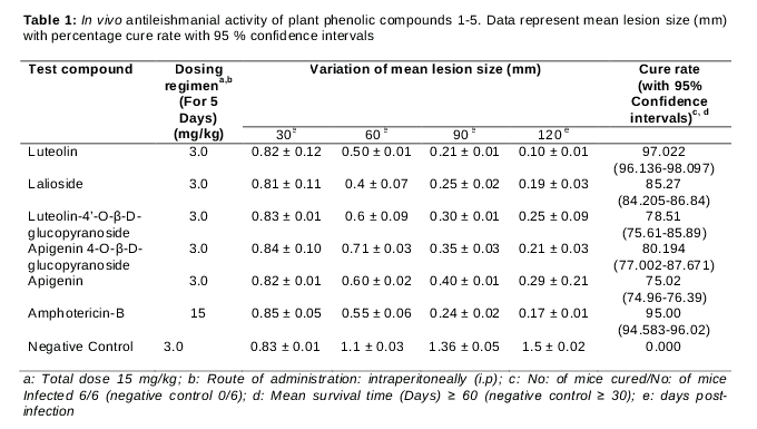
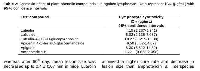
Results: For compound 1, mean lesion size decreased from 0.82 ± 0.12 to 0.10 ± 0.01 after 120 days, with 97 % cure of intracellular L. tropica amastigotes at a dose of 15 mg/kg, compared to amphotericin B which produced 95 % cure at a dose of 30 mg/kg. Half-maximal concentration (IC50) for compound 1 was 4.15 µg/ml against lymphocytes.
Conclusion: The results indicate that luteolin is a potent inhibitor of L. tropica amastigotes, with a higher cytotoxic activity against lymphocytes, compared with luteolin-4’-O-β-D-glucopyranoside.
Introduction
Leishmaniasis, caused by parasites belonging to the genus Leishmania (Family Trypanoso-matidae), is a major public health problem in tropical and sub-tropical regions. The parasite is transmitted by the sand fly (vector), with dogs, sheep, rats, horses, and cats being common animal hosts. The World Health Organization, WHO has reported that people from 98 countries in 5 continents, are at high risk of leishmaniasis, and it is estimated that approximately 12 million people are currently infected [1-3]. Cutaneous leishmaniasis is caused by different Leishmania species, e.g. Leishmania tropica, Leishmania major, Leishmania amazonensis, and Leishmania brazillensis. In Pakistan, L. tropica and L. major are the main causes of cutaneous leishmaniasis [4,5]. First line therapy for cutaneous leishmaniasis in Europe, Asia and Africa is pentavalent antimonials, i.e., sodium stibogluconate and meglumine antimoniate [2]. However, antimonials have severe side effects like myalgia, pancreatitis, cardiac arrhythmia, hepatitis, and drug accumulation in liver and spleen. Thus, there is an urgent need for new chemical entities for non-toxic and effective treatment of leishmaniasis [2,4].
Phenolic compounds comprise different groups including coumarins, flavonoids, tannins, naphthalenes, naphthoquinones, xanthones, lignans and alkylphenones [6]. Flavonoids and alkylphenones possess a variety of biological and pharmacological activities, including antioxidant [7], antibacterial, antiulcer [8], antifungal [9], antiviral [10], HIV - inhibiting [11], and anti-herpes simplex type 1 (HSV-1) [12]. They also inhibit electron transfer in mitochondrial inner membrane [13]. Flavonoids and alkylphenones are chemically well investigated, and a large number of member compounds have been reported. These include flavonoids and luteolin [14]; luteolin-4’-O-β-D-glucopyranoside [15]; apigenin 4-O-β-D-glucopyranoside [16]; apigenin [15]; xanhoangelol [8]; macaranggin [7]; artelastin [10]; 5,7-Dihydroxy-6,8-diphenyl flavonoids [11]; leachianone G [12] and alkylphenones such as Lalioside [17], 2,4,6-trihydroxyacetophenone-2-O-β-D-glucopyranoside [18] and lawsoniaside [19].
However, the current work deals with anti-leishmanial and cytotoxic effects of naturally occurring plant phenolic compounds having four flavonoids and one alkylphenone.
Methods
Chemicals
Fetal bovine serum, dimethylsulphoxide, Luteolin, Lalioside, luteolin-4’-O-β-D-glucopyra-noside, apigenin 4-O-β-D-glucopyranoside, apigenin and RPMI-1640 medium were purchased from Sigma-Aldrich (St. Louis, MO), whereas penicillin and streptomycin were purchased from MERCK, U.S.A. Amphotericin B was supplied by AmB-Fungizone, Bristol-Myers Squibb, UK. Water used for in vivo test solutions was purified by deionization and 0.22 µm membrane filtration (Millipore, Billerica, MA).
In vivo test
To study the pathogenesis of leishmania strain, 9 groups of male BALB/c mice (aged 6 - 8 weeks, and weighing 20 - 32 g) were used. Drug administration was through cardiac route. Promastigotes of L. Tropica KWH23 [21] were cultured in RPMI-1640 medium along with 10 % fetal bovine serum, penicillin (200 U/ml) and streptomycin (0.2 mg/ml). The parasite was cultured at 26 °C for 4 days in BOD incubator (Gallenkamp, Size 1, UK), and then harvested parasite. The harvested promastigotes were taken in a sterile tube and counted in a haemocytometer (REICHERT, N.Y, USA) under upright microscope (CX31, OLYMPUS, Tokyo, Japan). The promastigotes were centrifuged at 4 °C for 10 min at 2000 rpm; the supernatant liquid was discarded while pellet was left in tube. Fresh RPMI-1640 medium with 10 % FBS was added to get the required volume (10ml). The required volume (10 µl) of promastigotes (containing 1.4 x 10⁶ promastigotes/ml) was injected into the cardiac cavity (intraperitoneally) of the BALB/c mice. Developed lesions were measured weekly with dial micrometer (Mitutoyo, Japan) during the infection period. Infection was well established and clearly visible lesions were evident to the naked eye after 36 days. Then treatment process was started. Dose of test compounds given to Groups I, II, III, IV and V were 3.0 mg/kg for 5 days (total dose = 15 mg/kg) in DMSO up to final volume of 3 ml. Amphotericin-B was used as standard drug (positive control) at a dose of 15 mg/kg. No drug agent was used in VII group (negative control). The injection dose of 10 µl was given five times with 3-day intervals, and lesion measurements were recorded regularly. Dial micrometer was used to note the difference between size of the lesion in infected and uninfected mice weekly. Before and after treatment, needle aspirations were taken from the lesions [22]. To detect amastigotes under upright microscope, Giemsa stain was used, and the samples were examined under oil immersions. On the 30th, 60th, 90th and 120th day of infection, 60 mg of tissue sample was taken from the lesion for biopsy. In the identification of amastigotes, sample was smeared on the slides stained with Giemsa and upright microscope was used for examination.
Ethics statement
BALB/c mice were supplied by Department of Pharmacology (Animal center), University of Peshawar, KPK, Pakistan and this study was approved by Animal and Ethics Committee, Faculty of Pharmacy and Health Sciences, University of Balochistan (UOB), Quetta (approval ref. no. 093/FOPHS/UOB). The animals were maintained in accordance with UOB, Quetta Policy and international guidelines on the care and use of laboratory animals [20]. Standard diet along with water were given ad libitum to the BALB/c mice during experiments.
Cytotoxicity test
Fresh blood (10 ml) from a healthy volunteer was taken in BD vacutainer K2E (EDTA) to get mammalian cells (lymphocytes). Cytotoxic assay of test compounds was carried out by an adoption of the method described by Iqbal et al [22]. PBS was passed through 0.2 µm filter under laminar flow hood (kept sterile conditions) and then equal volumes of PBS and blood were taken in a sterile tube. Ficol solution (volume ratio 1:2) was carefully added at 165° angle to the mixture of PBS and blood. The tube was centrifuged at 2000 rpm at 4 °C for 30 minutes. The lower transparent portion was punctured with a syringe, and the liquid carefully removed and added to 5 ml of RPMI-1640 medium. The number of lymphocytes was counted in a haemocytometer under upright microscope. In the next step, 100 µl of the lymphocyte media was put into each well of a 96-well culture plate. The test compounds were added at doses of 100, 50, 25 and 10 µg/ml in DMSO, each in a final volume of 3 ml. Amphotericin-B (25 µg/ml, positive control) was used as reference drug, while negative control was L. tropica KWH23 promastigotes. Using a multipipette, 10 µl of promastigotes (containing 1.4 x 10⁶ promastigotes/ml) was added to 12 wells of the culture plate and placed in an incubator at 26 °C for at least 48 h. Haemocytometer was used to count viable lymphocytes and promastigotes under light microscope (lens 40x) at 24 and 48 hours. The cytotoxic tests were done in triplicate and the IC50 for each compound was calculated [23].
Statistical analysis
Results of in vivo anti-leishmanial assay of plant extract were expressed as mean % inhibition of parasite growth ± SD (n = 3). Cytotoxicity values were expressed as 50 % Inhibitory concentration (IC50) and analysed by non-linear regression analysis. For in vivo assays, mean lesion size (mm) and percentage cure were analysed by GraphPad Prism 5 software (GraphPad software, San Diego, CA) at 95 % confidence limit.
Results
Results of in vivo anti-leishmanial activities of compounds 1 - 5 analyzed in albino mice infected with 0.02 ml of clinically isolated L. tropica KWH23 having 1.4 x 10⁶ promastigotes, via intraperitoneal route, are shown . Mean lesion size decreased significantly from 0.29 ± 0.21 mm to 0.10 ± 0.01 mm after treatment with test compounds but that of the negative group reached 1.5 ± 0.02 mm, whereas it decreased in the Amphotericin (standard drug) group from 0.85 ± 0.05 mm to 0.17 ± 0.01mm after 120 days. Luteolin showed the strongest anti-leishmanial activity. The mice groups that received apigenin, luteolin-4’-O-β-D-glucopyranoside, and apigenin 4-O-β-D-glucopyranoside had mean lesion sizes of 0.29 ± 0.21 mm, 0.25 ± 0.09 mm and 0.21 ± 0.03 mm respectively, while corresponding percentage cure were 75.02, 78.51 and 80.19 % respectively. With Luteolin and lalioside mean lesion sizes were decreased to 0.10 ± 0.01 and 0.19 ± 0.03 mm, corresponding to 97.02 and 85.27 % cure, respectively.
Cytotoxic effect
Results of cytotoxic activities of compounds 1-5 are summarized in . Compounds 3, 4 and 5 showed highest cytotoxic activities 10.27, 9.50 and 8.30 µg/ml respectively. Compound 3 exhibited the greatest cytotoxicity against lymphocytes, with IC50 value of 10.27 µg/ml (95 % C.I = 6.215-15.38). Compound 1 gave the lowest cytotoxic activity (IC50 = 4.15 µg/ml; 95 % C.I = 2.287-5.941), whereas compound 2 showed IC50 value of 5.02 µg/ml (95 % C.I = 2.136-7.087). In comparison, amphotericin-B (standard drug) displayed IC50 of 1.072 µg/ml (95 % C.I = 0.823-2.358).
Discussion
Compounds 1-5 were selected due to their promising in vitro anti-leishmanial effects [22] against L. tropica. All tested compounds showed a cure rate between 97.02 and 75.02 % at 3 mg/kg concentration (for 5 days) whereas cytotoxic effects against lymphocytes in terms of IC50 values ranging between 4.15 and 10.27 µg/ml. All the mice were cured at or after 120th day of infection, when compounds 1 - 5 were given; whereas after 60th day, mean lesion size was decreased up to 0.4 ± 0.07 mm in mice. Luteolin achieved a higher cure rate and decrease in lesion size than amphotericin B. Interspecies variation in sensitivity against luteolin have been observed, e.g., it showed in vitro IC50 of 12.5 µM against L. donovani [27,28] and reduced splenic parasitic load up to 80 % when 3.5 mg/kg (twice a week for 1 month) was given to L. donovani infected hamsters [29].
Luteolin decreased cell viability of human hepatoma HepG2 cells, result in decrease in cytotoxic activity up to 41 % [24]. In this study, lalioside, apigenin, luteolin-4’-O-β-D-glucopyranoside, and apigenin 4-O-β-D-glucopyranoside showed significant anti-leishmanial properties. Similar results have been previously reported. Tasdemir reported that apigenin showed in vitro anti-leishmanial activity against L. donovani with IC50 values 1.9 µg/ml [28]. Apigenin showed cytotoxic activity at 8.30 µg/ml and induced hepatotoxicity at 100 and 200 mg/kg whereas it was non-toxic in 25 and 50 mg/kg [25]. In another study, apigenin decreased viability of human hepatoma HepG2 cells up to 42 % [24]. Apigenin 4-O-β-D-glucopyranoside showed cytotoxic activity at a concentration of 9.50 µg/ml and exhibited antitumor activity towards Hep G2, Hep 3B, MCF-7, A549 and MDA-MB-231 [26]. In the present study, lalioside and luteolin-4’-O-β-D-glucopyranoside showed greater cytotoxicity against lymphocytes, with IC50 values of 5.02 µg/ml (95 % CL = 2.136-7.087) and 10.27 µg/ml (95 % CI = 6.215-15.38) respectively, compared with amphotericin B (IC50 values = 1.072 µg/ml ;95 % CL = 0.823-2.358). The cytotoxic effects of these compounds may be due to the presence of phenolic hydroxyl groups [28]. Hydroxyl groups have affinity for proteins and may result in inhibition of microbial enzymes and plant NADH dehydrogenase [30,31]. It is evident that luteolin and luteolin-4’-O-β-D-glucopyranoside merit further investigations as suitable drug candidates against L. tropica.
Although different activities pertaining to the compounds studied are available in literature, the current study is the first report on the anti-leishmanial activities of compounds 2 – 4 and the first comprehensive in vivo study of cytotoxic activities of compounds 1-5 against L. tropica and mammalian cells.
Conclusion
Luteolin is a potent anti-leishmanial agent, but is cytotoxic against lymphocytes. Luteolin-4’-O-β-D-glucopyranoside possesses significant anti-leishmanial activity, and is least toxic against lymphocytes. Lalioside exert anti-leishmanial activity while apigenin 4-O-β-D-glucopyranoside and apigenin have a moderate inhibitory effect on intracellular amastigotes of L. tropica strain. Further studies on luteolin, luteolin-4’-O-β-D-glucopyranoside and other isolated compounds are necessary to investigate their mechanisms of action, specificity and structure-activity relationships.
Declarations
Acknowledgement
References
Archives


News Updates